Atkinson, J., & Braddick, O. (2013). Visual development. In P. D. Zelazo (Ed.), The oxford handbook of developmental psychology (Vol. 1, pp. 271–309). Oxford University Press.
Bucalo, P. (2015, October). Falcon belly dance. Youtube. Retrieved from
https://www.youtube.com/watch?v=JGArTWOJtXs
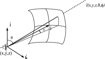
Chan, S. C. (2014). Plenoptic function. In K. Ikeuchi (Ed.),
Computer vision: A reference guide (pp. 618–623). Boston, MA: Springer US.
https://doi.org/10.1007/978-0-387-31439-6\_7
Charting, I. B. (2020a, June). Retinotopy task –
Ring-Expanding run. Youtube. Retrieved from
https://www.youtube.com/watch?v=DcgHJIlwQCo
Charting, I. B. (2020b, June). Retinotopy task –
Wedge-Clockwise run. Youtube. Retrieved from
https://www.youtube.com/watch?v=rsykP-9-moA
Edward H. Adelson, J. R. B. (1991). The plenoptic function and the elements of early vision. In
Computational models of visual processing. Retrieved from
http://citeseer.ist.psu.edu/viewdoc/summary?doi=10.1.1.2.9848
Gilmore, Rick O. (n.d.). Children’s brain responses to optic flow vary by pattern type and motion speed.
https://nyu.databrary.org/volume/75.
https://doi.org/10.17910/B7QG6W
Gilmore, R. O., Raudies, F., & Jayaraman, S. (2015). What accounts for developmental shifts in optic flow sensitivity? In
2015 joint IEEE international conference on development and learning and epigenetic robotics (ICDL-EpiRob) (pp. 19–25). ieeexplore.ieee.org.
https://doi.org/10.1109/DEVLRN.2015.7345450
Gilmore, R. O., Thomas, A. L., & Fesi, J. (2016). Children’s brain responses to optic flow vary by pattern type and motion speed.
PloS One,
11(6), e0157911.
https://doi.org/10.1371/journal.pone.0157911
Hofsten, O. von, Hofsten, C. von, Sulutvedt, U., Laeng, B., Brennen, T., & Magnussen, S. (2014). Simulating newborn face perception.
Journal of Vision,
14(13), 16.
https://doi.org/10.1167/14.13.16
Jandó, G., Mikó-Baráth, E., Markó, K., Hollódy, K., Török, B., & Kovacs, I. (2012). Early-onset binocularity in preterm infants reveals experience-dependent visual development in humans.
Proc. Natl. Acad. Sci. U. S. A.,
109(27), 11049–11052.
https://doi.org/10.1073/pnas.1203096109
Kiorpes, L. (2016). The puzzle of visual development: Behavior and neural limits.
J. Neurosci.,
36(45), 11384–11393.
https://doi.org/10.1523/JNEUROSCI.2937-16.2016
Koldewyn, K., Whitney, D., & Rivera, S. M. (2010). The psychophysics of visual motion and global form processing in autism.
Brain: A Journal of Neurology,
133(Pt 2), 599–610.
https://doi.org/10.1093/brain/awp272
Kretch, K. S., & Adolph, K. E. (2015). Active vision in passive locomotion: Real-world free viewing in infants and adults.
Developmental Science,
18(5), 736–750. Retrieved from
https://onlinelibrary.wiley.com/doi/abs/10.1111/desc.12251
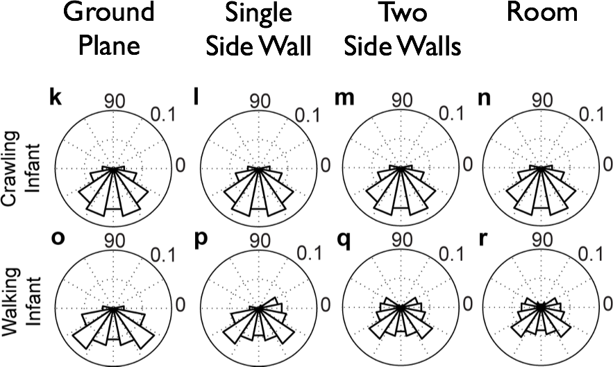
Kretch, K. S., Franchak, J. M., & Adolph, K. E. (2014). Crawling and walking infants see the world differently.
Child Development,
85(4), 1503–1518.
https://doi.org/10.1111/cdev.12206
Logothetis, N. K. (1999). Vision: A window on consciousness.
Scientific American,
281(5), 69–75. Retrieved from
https://www.ncbi.nlm.nih.gov/pubmed/10920769
Maurer, D., Lewis, T. L., Brent, H. P., & Levin, A. V. (1999). Rapid improvement in the acuity of infants after visual input.
Science,
286(5437), 108–110. Retrieved from
https://www.ncbi.nlm.nih.gov/pubmed/10506555
Mirabella, G., Kjaer, P. K., Norcia, A. M., Good, W. V., & Madan, A. (2006). Visual development in very low birth weight infants.
Pediatr. Res.,
60(4), 435–439.
https://doi.org/10.1203/01.pdr.0000238249.44088.2c
Qian, Y., Seisler, A. R., & Gilmore, R. O. (2021). Children’s perceptual sensitivity to optic flow-like visual motion differs from adults.
Developmental Psychology,
57(11), 1810–1821.
https://doi.org/10.1037/dev0001227
Randeberg, L. (2005). Diagnostic applications of diffuse reflectance spectroscopy. Retrieved from
https://www.semanticscholar.org/paper/ec9450b79923e2e2152b54ab9241b60bc5374944
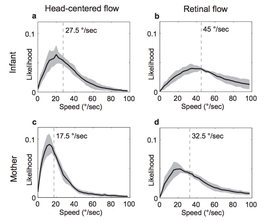
Raudies, F., & Gilmore, R. O. (2014). Visual motion priors differ for infants and mothers.
Neural Computation,
26(11), 2652–2668.
https://doi.org/10.1162/NECO\_a\_00645
Tootell, R. B. H., Tsao, D., & Vanduffel, W. (2003). Neuroimaging weighs in: Humans meet macaques in
“primate” visual cortex.
The Journal of Neuroscience: The Official Journal of the Society for Neuroscience,
23(10), 3981–3989.
https://doi.org/10.1523/JNEUROSCI.23-10-03981.2003